
Diagnostic Imaging
Imaging & specialist testing
St. George Hospital runs ultrasound, infrared thermography and darkfield microscopy on site as part of the diagnostic work-up, and arranges MRI, CT and PET-CT with partner facilities nearby when a question needs them. Your physician reads every image next to the laboratory results.
What imaging is available?
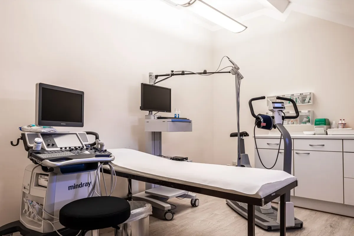
St. George Hospital runs three imaging and specialist techniques on site and arranges the rest. On site: ultrasound of the thyroid, abdominal organs, vessels and soft tissue; infrared thermography, which maps surface temperature patterns; and darkfield microscopy of a drop of live blood. For cross-sectional imaging, MRI, CT and PET-CT, the International Office books a partner facility nearby, usually within the first week. All of it is part of the diagnostic work-up and is read in the functional medicine consultation together with the laboratory profile.
The evidence differs by technique. Ultrasound is standard medicine with a large evidence base in its indications. Thermography has a thin evidence base and serves as a supplementary observation. Darkfield microscopy has no validated diagnostic accuracy; at most it prompts a laboratory test.
How each technique works
Ultrasound sends high-frequency sound into the tissue and builds an image from the echoes, in real time and without radiation. It shows the structure of soft tissue and organs, the size and texture of the thyroid, the liver, gallbladder, kidneys and spleen, fluid collections, and, with Doppler, the flow in arteries and veins. At the bedside it is part of the physician's examination. A 2025 appraisal of the point-of-care evidence found valuable sensitivity and specificity in many conditions, with the clearest value in biliary disease, ascites, causes of breathlessness and vascular questions, and noted that its effect on treatment decisions and outcomes is less well defined than its diagnostic accuracy (Ceriani et al., Eur J Intern Med, 2025).
The three on-site techniques compared
Ultrasound
- How it works
- Sound waves build a real-time image from the echoes, without radiation
- What it shows
- Thyroid, abdominal organs, soft tissue, fluid, and blood flow with Doppler
- Time and preparation
- 15 to 30 minutes on a couch; fasting for the abdomen
- Evidence
- Standard medicine with a large evidence base in its indications
Infrared thermography
- How it works
- A camera records skin temperature without contact
- What it shows
- Warmer and cooler regions: a pattern that shows where to look
- Time and preparation
- About 20 minutes in a room at a steady temperature
- Evidence
- Thin evidence base; a supplementary observation
Darkfield microscopy
- How it works
- A drop of fresh blood under a darkfield microscope, cells seen alive
- What it shows
- Red cell shape and clumping, white cell movement, plasma appearance
- Time and preparation
- One finger-prick drop; the image appears at once
- Evidence
- No validated diagnostic accuracy; may prompt a laboratory test
Your physician reads every finding together with the laboratory results. MRI, CT and PET-CT are booked with a partner facility.
What the imaging is used for
- Soft tissue and organ evaluation, abdominal organ screening
- Thyroid evaluation alongside the thyroid panel
- Vascular assessment with Doppler ultrasound
- Inflammatory focus detection, where thermography directs the ultrasound
- Musculoskeletal assessment before manual and physical therapies
- Anatomical evaluation before hyperthermia, apheresis or infusion therapies
- Staging and follow-up imaging in oncology, arranged with partner facilities and read with the treating oncologist
Which imaging do you still need?
Send the International Office a short history; the initial inquiry is free of charge. The Office then requests the scans and reports you already have for a medical review, which shows what we would image here, what we would book with a partner facility and what does not need repeating.
What a session looks like
Scheduling
Imaging is planned early. Abdominal ultrasound is done fasting; the other techniques need no preparation.
Examination
Ultrasound with gel on the skin while the physician explains the screen; thermography after a few minutes of acclimatization; one drop of blood for darkfield.
Discussion
All findings are discussed with you in the physician consultation, together with the laboratory results.
Your record
The images and reports are part of the written record you take home.
Evidence and safety
Ultrasound is strong in its indications and uses no radiation. Thermography is much less accurate than mammography for breast cancer screening, and darkfield microscopy has no validated diagnostic accuracy.
Show studies, risks and side effects
Ultrasound is among the most established imaging methods in medicine, with an evidence base across every specialty and no radiation. We grade it as strong in its indications.
Thermography is a different case. A systematic review of its use as a screening and diagnostic tool found a sensitivity of 25 percent for breast cancer screening compared with mammography, and diagnostic sensitivities ranging from 25 to 97 percent with specificities from 85 down to 12 percent, and concluded that there is not sufficient evidence to support thermography in breast cancer screening or as an adjunct to mammography (Fitzgerald et al., N Z Med J, 2012). A second systematic review of 60 studies reached the same conclusion for digital infrared thermal imaging, with sensitivity and specificity varying too widely between studies to pool (Vreugdenburg et al., Breast Cancer Res Treat, 2013). We grade thermography as limited and do not use it for cancer screening or diagnosis; its place here is as a temperature map that directs other examinations.
Darkfield microscopy has no diagnostic accuracy studies for any disease. It is used primarily in European integrative medicine, and we use it as a qualitative observation of blood morphology that is interpreted alongside standard laboratory testing, never as a stand-alone finding. What the evidence does not show is that either thermography or darkfield microscopy can establish or exclude a diagnosis.
Safety: ultrasound and thermography carry no known risk; darkfield microscopy involves a finger prick. MRI at a partner facility requires the usual screening for implants and metal, and CT and PET-CT involve ionizing radiation, which is why recent scans from home are read first and repeated only when the question requires it. All imaging is performed by qualified medical staff.
Costs: on-site ultrasound, thermography and darkfield microscopy appear in your written, itemized cost overview (package price plus individual add-ons). A deposit is paid before admission; treatment is self-pay and reimbursement is never promised. Costs and insurance
Frequently asked questions about imaging
Can you do an MRI or CT at the hospital?
Not on site. MRI, CT and PET-CT are performed at partner facilities nearby. The hospital books the appointment, usually within the first week of the stay, and arranges transport. The images and the radiologist's report come back to your attending physician and are discussed with you in the same consultation as the laboratory results. Recent scans from home are read first and are not repeated without reason.
Is thermography a cancer screening test?
No. Systematic reviews found that thermography's sensitivity for breast cancer is too low and too variable to use it for screening or to replace mammography, and we do not use it that way. Here it maps surface temperature patterns that can point to inflammation or circulatory change, as a supplementary observation read next to ultrasound and laboratory findings.
What does darkfield microscopy show?
A drop of fresh blood from a finger prick, viewed under a darkfield condenser so that cells appear bright against a dark background. It shows red cell shape, aggregation and the behavior of white cells in real time. No diagnostic accuracy study validates it for any disease, so it is an observation that may prompt a laboratory test, never a diagnosis on its own.
Do I need to prepare for ultrasound?
For an abdominal ultrasound, nothing to eat for about six hours so that the gallbladder is full and the bowel is quiet; water is fine. Thyroid, vascular and soft-tissue ultrasound need no preparation. The examination takes 15 to 30 minutes, is painless and involves no radiation; the physician explains what is on the screen as they go.
How much does imaging cost?
On-site ultrasound, thermography and darkfield microscopy are listed in the diagnostics block of your itemized estimate; MRI, CT or PET-CT at a partner facility are listed separately with the partner's fee. You receive the estimate in EUR after a physician has read your findings and before anything is booked. Treatment is self-pay; the invoice is itemized in English for out-of-network review.
References
- Point of care ultrasound: focus on evidence for a critical appraisal.. Eur J Intern Med 2025. PMID 40527717
- Thermography as a screening and diagnostic tool: a systematic review.. N Z Med J 2012. PMID 22426613
- A systematic review of elastography, electrical impedance scanning, and digital infrared thermography for breast cancer screening and diagnosis.. Breast Cancer Res Treat 2013. PMID 23288346
Send a free inquiry
The initial inquiry is free of charge. The International Office reviews your message and your case directly and gets back to you.
- Phone
- +49 8061 398-0
- Hours
- Mon–Thu 08:00–17:00, Fri 08:00–14:00 German time (CET/CEST); in the US Mon–Thu 2–11 a.m. ET, Fri 2–8 a.m. ET.
Munich Airport (MUC), about 60 minutes by car.